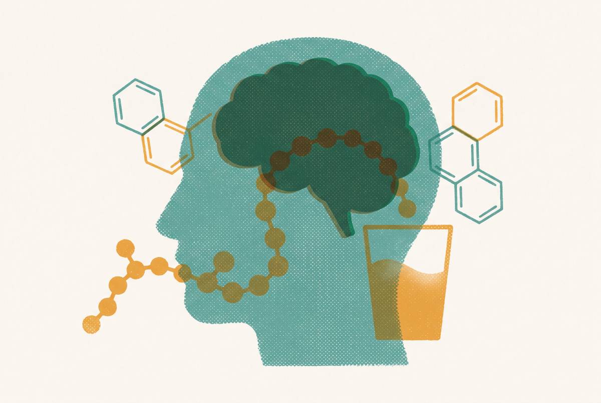
GLP-1 Receptor Distribution in the Brain and Gut
Mapping where GLP-1 receptors live in the brain and gut reveals what these drugs can actually do.
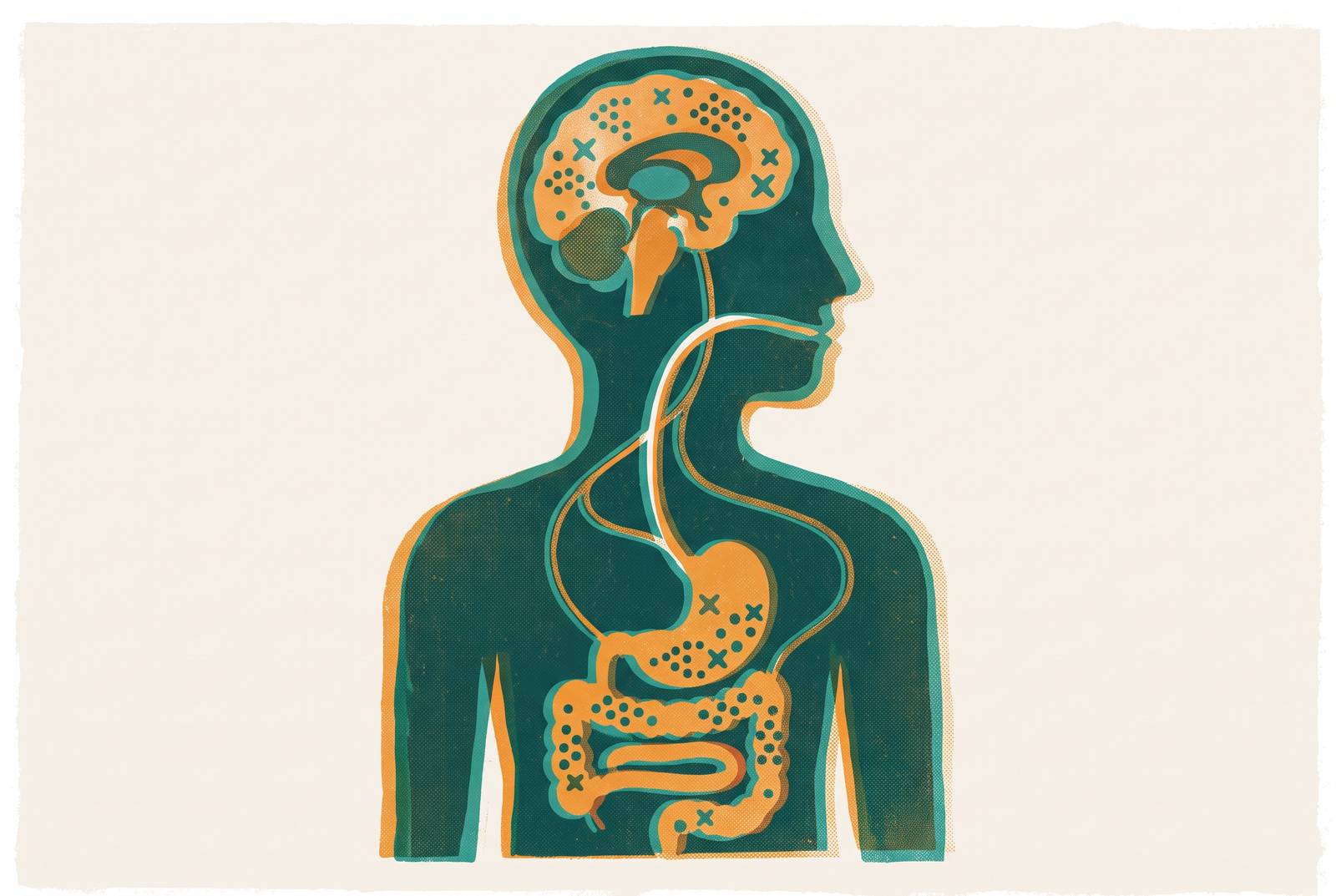
The GLP-1 receptor's tissue address determines its therapeutic biography. Where a receptor sits in the body dictates which cells it can influence, which circuits it can modulate, and which drugs can actually reach it, so mapping the receptor's distribution is the same project as mapping what GLP-1 medicines can and cannot do. The receptor belongs to the class B1 GPCR family, built to recognize peptide hormones rather than small molecules, and its large extracellular domain captures the peptide's C-terminus while the N-terminus inserts into the transmembrane bundle to trigger activation. Drug Discovery News described this two-domain binding mechanism in June 2026, along with the sharp kink that forms in transmembrane helix 6 once the peptide docks, a structural pivot that swings the receptor's intracellular half outward and opens a face for the Gs protein to bind. That pivot is the molecular event that begins every GLP-1 signal, in the pancreas, the gut, or the brain.
From there, the receptor signals primarily by raising cyclic AMP through adenylyl cyclase, though it also recruits beta-arrestins. That dual signaling creates a balance between G-protein activation and arrestin recruitment, a phenomenon called biased agonism, now an active target of drug design. What matters just as much as the signaling chemistry is where the receptor actually sits. Validated antibodies and reporter models have corrected a set of earlier over-attributions, showing that some tissues once assumed to express GLP-1R do not. Therapeutic assumptions built on those older, unvalidated maps have had to be revised. The confirmed distribution reaches pancreatic beta cells, the sinoatrial node of the heart, vascular smooth muscle, the enteric nervous system, and several appetite and reward centers in the brain, a footprint that extends the receptor's relevance well past the pancreas and into cardiovascular, digestive, and neurological territory.
How the gut produces GLP-1
The gut is where GLP-1 is made, and the receptor population there is functionally real, but peripheral signaling was never built to be the only channel to the brain. GLP-1 originates in L cells, a sparse population of enteroendocrine cells concentrated in the distal small intestine and colon, with a smaller contingent in the proximal small intestine that may explain how quickly GLP-1 levels rise after a meal. A 2026 review by Beutler proposed that these proximal L cells work alongside a neuroendocrine feedback loop to accelerate that post-prandial release.
GLP-1R itself is expressed throughout the human enteric nervous system, with higher expression in the colon than in the stomach or ileum, concentrated particularly in the distal colon. The receptor is not spread evenly across gut tissue. It localizes selectively to distinct neuron subtypes, which tells a more specific story than diffuse hormone action: certain enteric neurons are built to respond to GLP-1 and others are not. Once GLP-1 is released, it acts on vagal afferents, the nerve fibers that relay information from the abdomen up to the brainstem. That relay is the gut-to-brain connection most people picture when they think about GLP-1 and appetite, but it works indirectly. The gut does not send the hormone itself to the brain, it sends a neural signal carried by the vagus nerve.
The reason that distinction carries weight is that native GLP-1 barely survives outside the gut. Circulating GLP-1 gets broken down by the enzyme DPP-4 within roughly two minutes of release. That two-minute window drove essentially all of the pharmaceutical engineering that followed: acylation, fatty-acid tethering, and SNAC chemistry all exist to extend a molecule's half-life long enough for it to survive in plasma. Seen from that angle, the entire project of engineering longer-acting GLP-1 drugs is a workaround for a signal that was never meant to travel through the bloodstream to reach the brain in the first place. The gut's native architecture sends a fast, local, self-limiting signal through nerves rather than a durable hormone through circulation.
This has direct consequences for what the peripheral receptor population actually accomplishes. The incretin effect, the glucose-lowering action that GLP-1 is best known for clinically, is mediated largely by receptors on pancreatic beta cells. Conditional knockout studies have confirmed as much: beta-cell GLP-1R, not receptors in the brain or the peripheral nervous system, carries primary responsibility for the incretin and glucose-lowering effects of GLP-1 receptor agonists. That finding draws a sharp line through the receptor map. Metabolic glucose control lives in the gut and pancreas. Weight loss and any neurological effect have to be explained by receptors somewhere else, which raises an immediate question: where in the brain does that somewhere else actually sit?
The brain's own GLP-1 system: NTS neurons, PPG projections, and central receptor populations
The brain does not simply wait for gut-derived signals to arrive. It manufactures its own GLP-1, through a dedicated population of neurons whose output pattern lines up almost precisely with where GLP-1 receptors sit in the central nervous system. That alignment amounts to an intrinsic delivery infrastructure the brain built for itself, independent of anything happening in the intestine.
The source of that central supply is a group of neurons called preproglucagon neurons, or PPG neurons, located in the nucleus of the solitary tract (NTS) in the caudal brainstem. Retrograde tracing studies have also shown that NTS PPG neurons project directly to the nucleus accumbens, tying the brainstem's GLP-1 system to the mesolimbic reward circuit by a direct anatomical route. The distribution of PPG axon terminals mirrors the distribution of GLP-1 receptor cells across the CNS, so the brain's own release sites and its own receptor populations are co-located by design.
PPG neurons do not confine themselves to a single feedback loop. They project broadly to autonomic control sites throughout the brain, functioning as a distributed modulatory system woven into energy balance, stress response, and reward rather than a single local circuit. Brain-derived GLP-1 has a demonstrated role in suppressing both hedonic food intake, eating driven by pleasure rather than need, and metabolic food intake, eating driven by caloric requirement. The NTS is a critical hub for integrating peripheral nutritional signals from the gut: it receives vagal afferent input carrying signals from the gut, and it also produces its own GLP-1, making it a convergence point for both the peripheral relay and central GLP-1 generation.
How much gut-derived GLP-1 ever reaches these central receptor populations directly, versus how much of the brain's response depends on its own supply, remains genuinely unresolved. That uncertainty is itself an argument for why central receptor targeting deserves attention on its own terms, rather than being treated as a downstream extension of gut pharmacology. If the brain runs its own parallel system, then reaching that system directly, rather than hoping a peripheral signal eventually gets there, becomes a design question in its own right.
Hypothalamic and reward-circuit pathways for GLP-1 receptor activation and reduced intake
GLP-1 receptor agonist efficacy in obesity traces back to activation of receptors inside the central nervous system, not to the peripheral incretin action described earlier. The brain's receptor geography is the mechanism behind the drugs' most consequential effect on body weight. Inside the arcuate nucleus of the hypothalamus, GLP-1R appears on two distinct neuron populations that pull in opposite directions. It sits on POMC neurons, which suppress appetite, and on neurons expressing thyrotropin-releasing hormone, which send inhibitory signals to AgRP neurons, the cells that drive hunger. The receptor therefore occupies the intersection of both arms of the hypothalamic circuit that governs energy balance. Which of these populations matters most for producing benefit, and which contributes to unwanted effects, remains an open question. Beutler's 2026 review frames identifying the specific GLP-1R-expressing cells responsible for each outcome as an active and unresolved research question driving next-generation drug design.
One might argue that the mechanism is already well enough understood to stop asking these questions, but a 2026 paper in Nature suggests otherwise. Researchers used humanized GLP-1R mouse models to trace the neural circuitry through which small-molecule GLP-1R agonists modulate feeding, work that carries direct implications for how orally delivered weight-loss drugs engage brain reward circuits. That circuitry extends into mesolimbic pathways, including the nucleus accumbens, and activation there appears to explain something patients describe often but clinicians have struggled to name precisely: a quieting of what's commonly called "food noise," the constant background pull toward eating. That effect looks less like simple appetite suppression and more like a reduction in the motivational salience food carries, a dampening of how strongly food-related cues compete for attention. The Physiologist Magazine reported that researchers are tracing GLP-1 agonist influence across inflammation, cardiovascular health, mood, motivation, and reward, extending well past the downstream effects of weight loss alone.
But what if the same receptor populations producing these benefits are also producing the drugs' most common complaint? Recent data indicate exactly that: the aversive gastrointestinal side effects associated with GLP-1RAs, nausea and vomiting chief among them, trace back to CNS-mediated activity in the same receptor populations responsible for appetite suppression. With 40 to 60 percent of GLP-1 users reporting GI side effects in clinical trials, that overlap ties the discomfort to the same receptor populations that produce the benefit. It is a brain-receptor targeting problem, since the same central receptors that produce the benefit also produce the discomfort, so how a drug reaches those receptors, and how selectively, becomes the whole game.
Why current delivery routes reach brain GLP-1 receptors inefficiently
Current subcutaneous and oral GLP-1 receptor agonists push plasma concentrations high, yet achieve only limited direct exposure inside the brain. That mismatch between how much drug circulates peripherally and how much reaches central receptor populations explains both the side-effect burden described above and the ceiling on how far these drugs can extend into neurological indications. Subcutaneous injection remains the dominant format by revenue: market data reported by Yahoo Finance show injections represented the large majority of GLP-1 revenue in 2025. That dominance reflects commercial momentum built around an established delivery method, not necessarily the optimal route for reaching brain receptor populations. The blood-brain barrier remains a genuine pharmacological obstacle. Trials aimed at neurological indications have produced mixed results, and poor penetration across that barrier has been identified as a causal factor in several of them.
The clearest evidence of this delivery gap comes from a pair of Alzheimer's disease trials. Two Phase 3 trials of oral semaglutide in early Alzheimer's disease, each enrolling thousands of participants, failed to significantly slow disease progression. Around the same period, the ELAD trial, a randomized, double-blind, placebo-controlled study of patients with mild Alzheimer's disease, tested injectable liraglutide and found roughly 50 percent less brain volume loss across frontal, temporal, parietal, and total gray matter regions compared to placebo. That finding was an exploratory endpoint rather than the trial's primary measure, and the primary endpoint, cerebral glucose metabolic rate, was not met. The result should be read cautiously rather than as confirmation of anything definitive. The pattern suggests the biological target may be valid even where a given formulation fails to reach it.
The GI side effects reported across a large share of trial participants represent the downstream cost of this same mismatch. Reaching any meaningful concentration of drug inside the CNS currently requires systemic levels high enough to also saturate peripheral GLP-1 receptors, in a way the enteric nervous system was never built to tolerate at pharmacologic doses. Patients who stop treatment because of side effects or needle burden are not failing to comply with a workable regimen. The regimen itself is asking a peripherally dosed drug to do a job that depends on reaching a receptor population sitting behind the blood-brain barrier, and current routes accomplish that inefficiently. The receptor map has identified real, valid targets throughout the CNS. What has not caught up is a delivery method built to reach them without saturating everything else along the way.
The reward circuitry GLP-1R population and addiction as the neurological indication with the strongest current evidence
GLP-1 receptors positioned inside the mesolimbic and mesocortical pathways are not incidental outposts of a peripheral hormone system. They function as core nodes within the reward circuitry itself. That makes addiction the neurological indication most directly explained by the receptor's own distribution. The same pathways responsible for quieting food noise in obesity also appear to mediate craving in substance use disorders, since GLP-1 receptors sit within the reward circuits that govern addictive behavior broadly, not merely appetite-driven behavior. Preclinical studies have shown that GLP-1 agonists consistently reduce voluntary self-administration of alcohol, cocaine, fentanyl, heroin, and nicotine across animal models, apparently by altering the underlying neurobiology of the reward circuit rather than by any peripheral mechanism. A study published in The Lancet offered the most compelling clinical evidence gathered so far that GLP-1 receptor agonists can treat addiction in humans.
What makes this application mechanistically coherent, rather than a fortunate side effect discovered by accident, traces back to the anatomy covered earlier. The nucleus accumbens receives direct PPG neuron projections originating in the NTS. The brain's own GLP-1 system was wired into the reward circuit long before any drug was designed to act on it. A pharmaceutical that engages this system is not creating a connection so much as exploiting one the brain already built.
That raises an important question, though, and it deserves a direct answer rather than a dismissal. If injectable GLP-1RAs already reach reward circuits well enough to reduce substance use in the studies so far, why does delivery route still matter here? The addiction evidence, promising as it looks, remains largely observational and preclinical rather than the product of large confirmatory trials. The dose required to reliably engage reward-circuit GLP-1 receptors through systemic injection may differ meaningfully from what the NTS-PPG system delivers locally and continuously as part of normal brain function. Side effects at doses sufficient to achieve that engagement remain a genuine barrier to population-scale use in addiction treatment, a population that may tolerate gastrointestinal discomfort and injection burden less readily than patients pursuing weight loss.
None of that undercuts the underlying anatomy. The receptor map across gut, brainstem, hypothalamus, and reward circuitry keeps identifying targets worth pursuing, and each new application makes the same point from a different angle. Knowing where a receptor sits explains what a drug can plausibly do. Getting a drug there cleanly, without saturating every other receptor population along the way, remains the unfinished part of the work.
Sources
- GLP-1 physiology and pharmacology along the gut-brain axis - PMC
- GLP-1 Drugs and the Physiology of Metabolic Health
- GLP-1 receptor biology: structure, signaling, and distribution
- A brain reward circuit inhibited by next-generation weight-loss drugs in mice
- Distribution and ultrastructural localization of the glucagon-like peptide-1 receptor (GLP-1R) in the rat brain
- Systematic Characterisation of GLP‐1R in Human Enteric Nervous System: Implications for GLP‐1 as a Key Regulator of Colonic Activity - PMC
- GLP-1 Neurons in the Nucleus of the Solitary Tract Project Directly to the Ventral Tegmental Area and Nucleus Accumbens to Control for Food Intake - PMC
- Brainstem GLP-1 neurons modulate physiological satiation and drive sustained weight loss in obese mice - PMC