GLP-1 Peptide Degradation Pathways and Stability Challenges
Blocking three enzymes may be needed to keep GLP-1 stable enough for therapy.
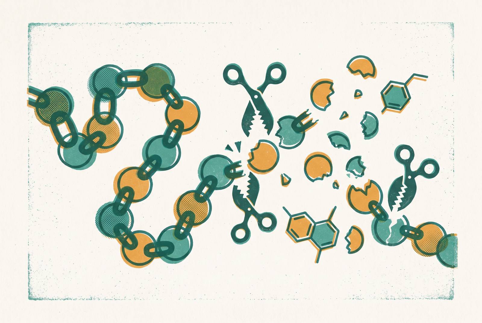
GLP-1's trouble as a drug candidate starts at the moment of its birth. The hormone is released by intestinal L-cells in response to a meal, and its two bioactive forms, GLP-1(7-36)NH₂ and GLP-1(7-37), both bind and activate the class B G-protein-coupled GLP-1 receptor. That activation is what drives glucose-dependent insulin secretion, suppresses glucagon, slows gastric emptying, and shapes appetite. But the body never built GLP-1 to last. The body built GLP-1 to signal once, briefly, after food arrives, and then disappear, and that disappearance is the design.
Within minutes of release, the peptide runs into dipeptidyl peptidase-4, an enzyme that starts clipping it almost as fast as the L-cells can secrete it. Peptide that survives this first pass does not get a clear road after that. The hepatic bed supplies a second clearance opportunity, where intact GLP-1 and the related hormone GIP face further inactivation by DPP-4 bound to hepatocytes as blood moves through the liver. Degradation has already happened across three separate organs rather than at a single checkpoint by the time the kidney finishes eliminating the resulting metabolites. Gut, liver, kidney: each one takes a turn.
The practical consequence shows in what actually circulates. What reaches peripheral tissues is mostly the truncated, weakly active fragment GLP-1(9-36)NH₂, not the hormone that left the L-cell. So even before anyone asks how to formulate GLP-1 into a usable drug, a more basic fact is already working against the effort: the native molecule in circulation is already mostly a degradation product. Its natural role is a short postprandial signal, and that short signal is why therapeutic deployment runs into problems from the first step. Every strategy discussed in the sections that follow, whether it targets an enzyme, a filtration mechanism, or a formulation weakness, exists because this baseline biology leaves so little room to work with. DPP-4 moves first and fastest, but it is not acting alone, and the rest of this piece maps out who else is involved.
How DPP-4 disables GLP-1 at the N-terminus
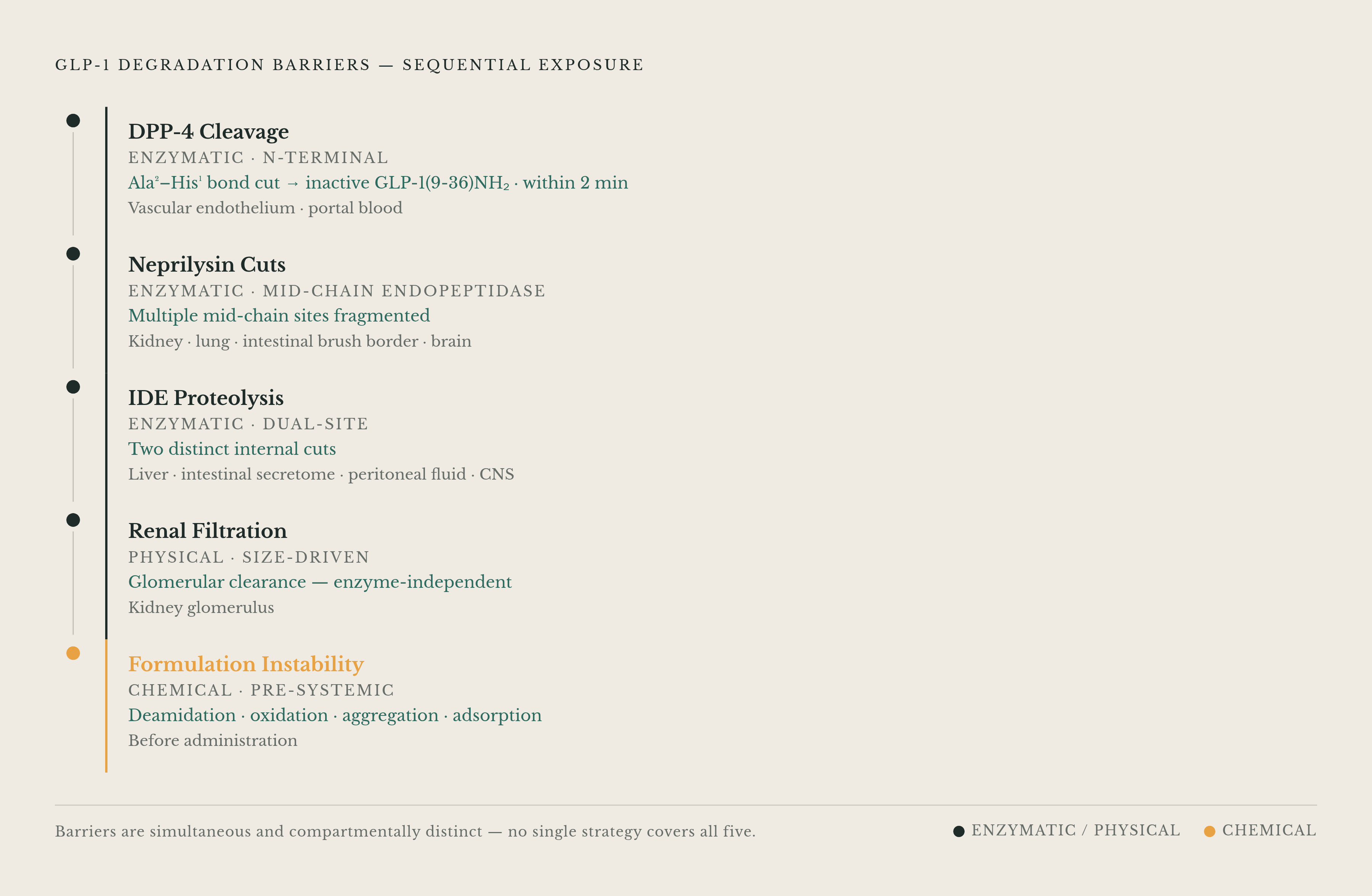
DPP-4 does not chew through GLP-1 indiscriminately. It makes one precise cut, at the Ala²-His¹ bond near the peptide's N-terminus, and that single break is enough to take the molecule out of play. In full proglucagon numbering, this is the Ala8-Glu9 bond, and the serine protease that performs the cleavage converts active GLP-1(7-36)NH₂ into GLP-1(9-36)NH₂, the form that then dominates circulation. One bond, one enzyme, and a hormone's signaling capacity is gone.
That narrowness in where DPP-4 attacks is also what makes the attack easy to design around, at least in principle. Because the enzyme needs a specific structural target at a specific position, researchers can intervene at that exact spot. Substituting the amino acid at position 2, using building blocks such as Aib or D-amino acids, became the earliest and still the most widely used way to block DPP-4 from getting its grip. Structural biology has guided this work closely, using the known geometry of the cleavage site to inform which substitutions at which positions hold up against the enzyme without destroying the peptide's other functions.
That last qualifier carries real weight. The GLP-1 receptor itself needs an intact N-terminus to achieve full agonism, so any change made to dodge DPP-4 has to leave the receptor-binding geometry close to untouched. Engineers are threading a needle: alter the region enough to block the protease, but not so much that the receptor stops recognizing the peptide as a signal worth answering. And DPP-4 is not a narrow threat confined to one compartment. DPP-4 is present in plasma, on endothelial surfaces, and on immune cells, so resistance engineered against it has to hold up everywhere the enzyme is present.
Solving the N-terminus problem, though, only solves the N-terminus problem. The rest of the peptide chain remains exposed, and that is where the next enzyme comes in.
What neprilysin does to the fragments DPP-4 leaves behind
Even a GLP-1 analogue that shrugs off DPP-4 completely still has to get past neprilysin, also called NEP 24.11, an endopeptidase that cuts the peptide at multiple points along its mid-chain. NEP does not care whether the peptide is the original active hormone or the truncated product DPP-4 already produced. It degrades both GLP-1(7-36)NH₂ and GLP-1(9-36)NH₂, breaking each into smaller fragments that carry no receptor activity.
Direct measurement showed how much this enzyme actually matters. A 2025 study published in the Journal of the Endocrine Society tested what happens when both DPP-4 and neprilysin are inhibited at once, comparing the resulting plasma levels of intact GLP-1 against a condition where neither enzyme is blocked. Blocking both produced a far larger area under the curve for intact, circulating GLP-1 than blocking neither. That gap matters because it shows neprilysin is a co-primary degradation pathway, operating on its own timescale and contributing its own share of the peptide's disappearance.
The design consequence follows directly. An analogue engineered only for DPP-4 resistance still carries mid-chain residues that neprilysin can reach and cut, so the half-life gained from N-terminal protection alone is partial at best, not complete. Real durability requires addressing both enzymes, and that requirement only grows once the anatomy of neprilysin's reach is taken into account. The enzyme is expressed in the kidney, the lung, the intestinal brush border, and the brain. Its relevance extends past plasma pharmacokinetics into tissue compartments, including the brain, that become central once delivery routes beyond standard injection enter the conversation.
So the degradation map, at this point, already has two enzymes working in sequence on circulating peptide. A reader might reasonably assume that covers the major threats, but it does not.
The third degradation actor: insulin-degrading enzyme in tissue and the CNS
A third enzyme enters the picture, and it is the one least likely to be on anyone's radar going into this conversation. Insulin-degrading enzyme, or IDE, was identified in a 2026 study published in Science Advances by researchers at the Shanghai Institute of Organic Chemistry, Chinese Academy of Sciences, as a previously unrecognized protease acting on GLP-1, cutting the peptide at two distinct sites. The study's authors describe IDE-mediated degradation as a major mechanism regulating glucose control, placing IDE on comparable footing with DPP-4 and neprilysin.
What makes IDE's discovery change the shape of the whole problem is where that enzyme operates, not just that a third enzyme exists. IDE is tissue-resident, active in the liver, the intestinal secretome, peritoneal fluid, and the central nervous system. Measurements of circulatory half-life, the kind of data that has guided GLP-1 engineering for years, simply do not capture degradation happening in those compartments. A peptide can look stable in plasma and still be getting cut apart inside tissue the blood tests never examine.
The same Science Advances study tested a response: engineering GLP-1 and an existing analogue, Semaglutide, with D-amino acid substitutions placed at the IDE cleavage sites. The resulting peptides showed improved stability specifically in plasma, liver, intestinal secretomes, peritoneal fluid, and the CNS, which confirms both that IDE's threat is real across those compartments and that targeted substitution can address it there too.
The CNS piece of this carries particular weight. If IDE is active in the brain, then GLP-1 peptides arriving there by any route, intranasal delivery included, face a tissue-level degradation threat that fatty acid acylation and N-terminal modification were never built to stop. Those two strategies were developed against DPP-4 and against renal clearance, not against a protease operating inside brain tissue. The discovery of IDE does not just add a third item to a list. It reframes the entire problem: degradation is no longer just something that happens to circulating peptide over time, it is something that happens differently across several anatomical compartments, on several different timescales, and a solution built for one compartment carries no guarantee of working in another.
Renal clearance as the fourth constraint: size-driven elimination independent of enzymes
Three enzymes aside, a fourth problem remains, one that has nothing to do with enzymatic activity. GLP-1 is small enough to pass freely through the glomerulus, so the kidney filters it out of circulation regardless of whether DPP-4, neprilysin, or IDE have touched it. This clearance route runs in parallel with enzymatic degradation rather than depending on it, and resistance engineered against any or all three enzymes does nothing to slow it down.
The kidney's role here extends past native peptide. It also carries out final elimination of the metabolites that DPP-4 and neprilysin produce upstream, so renal clearance shortens overall half-life even in cases where enzymatic degradation has already been partly addressed through protein engineering. Resistance to the enzymes does nothing to stop the kidney from filtering the peptide out.
This is the mechanism behind why molecular enlargement became such a central engineering strategy. Fatty acid acylation that enables albumin binding, Fc fusion, and PEGylation all extend half-life largely by increasing the hydrodynamic radius of the molecule past the size the kidney can filter, rather than primarily by blocking enzymes from reaching their cleavage sites. Acylation, PEGylation, Fc fusion, and albumin fusion are all strategies that respond to the combined pressure of rapid DPP-4 cleavage and renal elimination acting together, not to either threat alone.
That solution, though, creates its own complication further down the line. A peptide built large and lipidated enough to escape renal filtration carries size and lipophilicity that make mucosal absorption and CNS entry harder, which matters considerably for any delivery route other than injection. Escaping renal filtration makes delivery by routes other than injection harder. Four mechanisms now sit on the table, three enzymatic and one physical, and the next question is what happens to a peptide engineered to resist all four before it ever gets into the body.
Formulation's fifth layer of instability
Everything described so far happens to GLP-1 inside a living system. A fifth category of instability happens before the peptide ever gets there, inside a vial, a syringe, a manufacturing line, or a shipping container, and the structural modifications built to fight enzymes and filtration tend to make this category worse.
Peptide therapeutics face chemical breakdown routes that small-molecule drugs mostly avoid. Asparagine and glutamine residues deamidate, a reaction that runs particularly fast at Asn-Gly sequences under ordinary physiological pH and temperature. Methionine and histidine residues oxidize when reactive oxygen species are present. Aspartate-proline bonds hydrolyze on their own. This It is chemistry acting on the peptide bond by bond, given enough time, heat, or exposure.
Physical instability layers on top of the chemical kind. Lipidated GLP-1 analogues, the same ones engineered for albumin binding and renal persistence, self-associate through hydrophobic interactions and form aggregates that cut bioavailability and raise the risk of an immune response. Under stress conditions, some peptides misfold into beta-sheet-rich amyloid fibrils. Even peptide that avoids both of those fates can still adsorb onto the glass, rubber, or plastic surfaces of its own storage container, quietly reducing the dose that actually reaches the patient. The same fatty acid chain that extends circulatory half-life by enabling albumin binding is also the chemistry that drives aggregation, so the modification solving one problem is directly responsible for creating another.
Temperature makes the picture less forgiving still. Freeze-thaw cycles, agitation during shipping, and ordinary temperature excursions unfold a peptide's secondary structure, and an unfolded peptide degrades chemically much faster than a folded one. That is why cold-chain dependency follows so directly from the molecule's chemistry, and why it becomes a real constraint on distribution and access anywhere reliable refrigeration cannot be guaranteed. Oral delivery adds yet another obstacle on top of all this: gastric acid, pancreatic proteases, and intestinal brush-border enzymes form an additional multi-enzyme gauntlet that even structurally modified peptides often fail to cross intact. In vivo degradation and formulation instability do not operate as separate problems to be solved one after another. They operate at the same time, on the same molecule, and that simultaneity is what makes the engineering challenge as difficult as it is.
What structural engineering has achieved against these pathways
Measured against the five fronts just described, structural modification of the peptide itself counts as a genuine achievement. N-terminal substitution, fatty acid acylation, and targeted amino acid engineering together turned a hormone that barely survives a few minutes in circulation into a peptide stable enough for once-weekly injection. That is not a small result. It represents a real answer to DPP-4 cleavage and, through the mechanism of albumin binding, a real answer to renal filtration as well, since longer and more complex acyl chains extend hydrodynamic radius further and push the half-life gain out even more.
But what has that engineering actually reached, and what has it left standing? The gains land squarely at the circulatory boundary. They slow DPP-4, and through molecular enlargement they outrun the kidney's filtration threshold. They do not, on their own, reach IDE sitting in the liver, the intestinal secretome, peritoneal fluid, or the CNS, compartments where circulatory half-life measurements never looked. They do not resolve the formulation instability that acylation itself introduces through aggregation and cold-chain sensitivity. And they do not address the degradation environment a peptide meets once it tries to reach the brain by a route other than the bloodstream.
None of that diminishes what structural modification accomplished against the threats it was built to answer. It does mean the degradation map laid out across this piece, DPP-4, neprilysin, IDE, renal clearance, and formulation instability, is wider than any single engineering strategy yet covers. Each piece of that map demands its own kind of intervention, and the five do not resolve into one unified fix. Understanding where the current engineering stops is what turns this from a tidy success story into the actual starting point for the next generation of GLP-1 delivery work.
Sources
- IDE-mediated GLP-1 degradation as the basis for designing long-acting and CNS-stable GLP-1 receptor agonists - PMC
- In Vivo Inhibition of Dipeptidyl Peptidase 4 and Neprilysin Activity Enables Measurement of GLP-1 Secretion in Male Rats
- Rational Design by Structural Biology of Industrializable, Long-Acting Antihyperglycemic GLP-1 Receptor Agonists