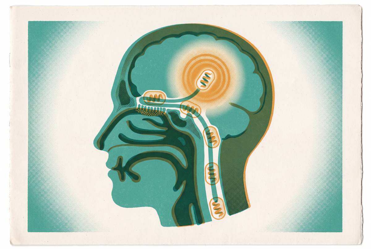
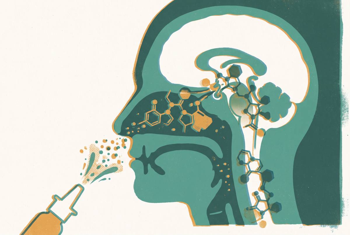
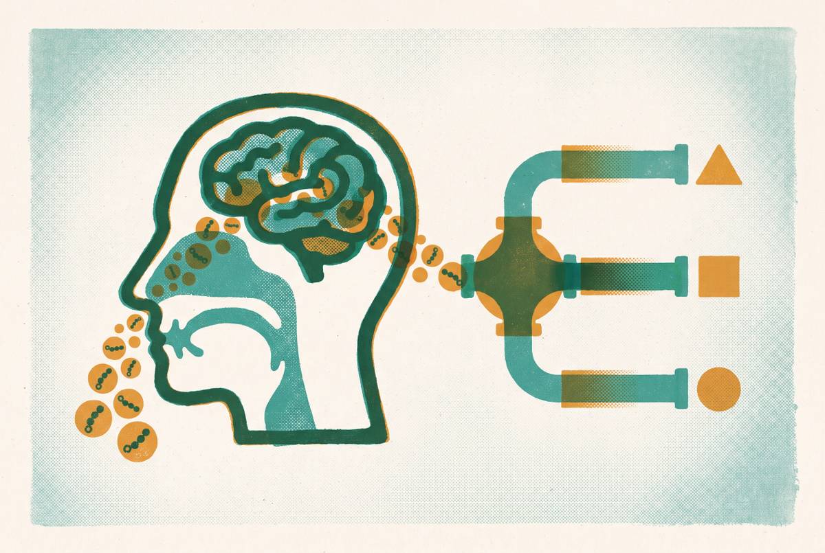
Olfactory Epithelium Anatomy and Drug Transport Pathways
Only one nasal tissue zone links to the brain, which shapes formulation.
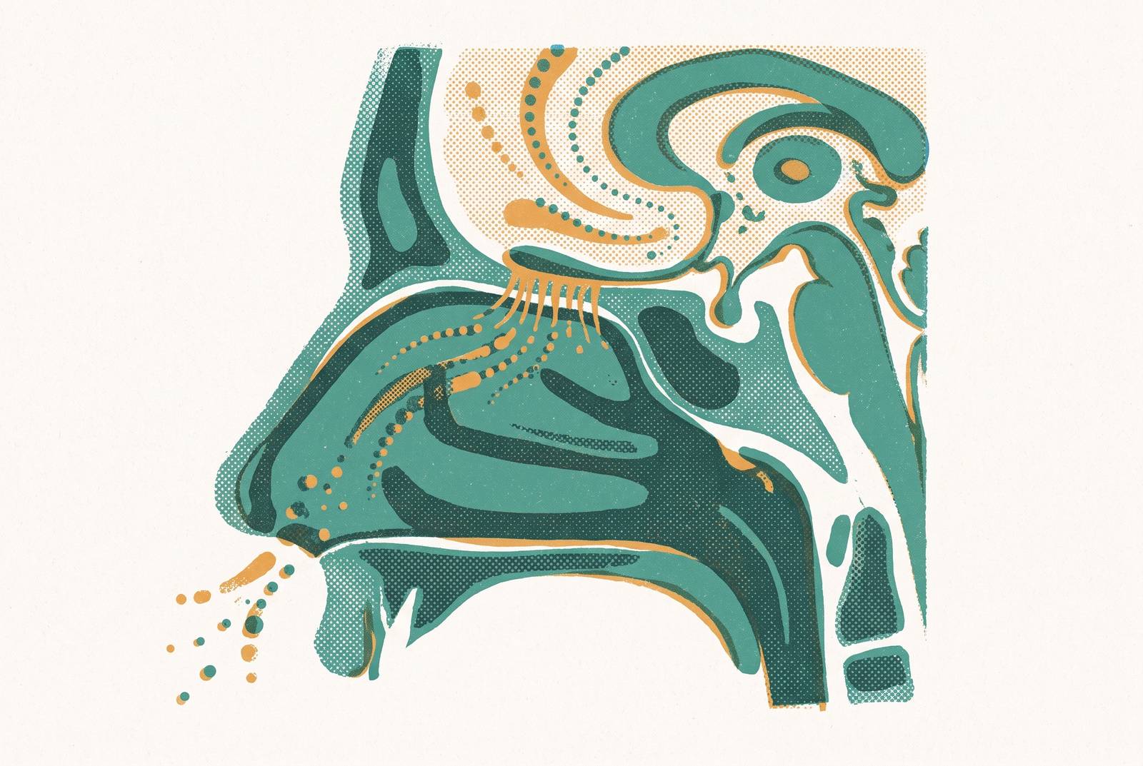
Most discussions of intranasal drug delivery treat the nasal cavity as a single absorption surface. The clinical assumption is that anything deposited in the nose has a chance of reaching the brain. The anatomy says otherwise. The nasal cavity contains two functionally distinct tissue zones with different cellular architecture, different innervation, and different transport capabilities. Only one of them connects directly to the central nervous system.
The olfactory epithelium is the only nasal tissue that gives direct brain transport. Its sensory neurons run uninterrupted from the mucosa through the cribriform plate to the olfactory bulb, so a drug can travel along them or through the perineural spaces around them and bypass the blood-brain barrier. The respiratory epithelium covers most of the nasal cavity and mainly supports systemic absorption, with CNS access only afterward via trigeminal nerves. A drug deposited there enters the bloodstream first and still has to cross the BBB, so the result is a different pharmacokinetic path and not a weaker version of direct delivery. In humans the olfactory zone is only about 10% of nasal surface area, which is why standard sprays tend to land in the wrong place.
The respiratory epithelium covers most of the nasal cavity. It is a protective surface, built to humidify and warm inhaled air and to trap and remove particles before they reach the lungs. The olfactory epithelium behaves differently. (2026) state the distinction plainly: only the olfactory epithelium enables direct brain transport through olfactory neurons, bypassing the blood–brain barrier, while the respiratory epithelium primarily supports systemic absorption before CNS access via trigeminal nerves.
The practical consequence matters for every intranasal formulation in development. Depositing a drug in the wrong nasal zone does not give a weaker version of direct brain delivery. It gives an entirely different pharmacokinetic path, with the BBB still in play.
What makes the olfactory epithelium different from the respiratory epithelium for direct brain transport?
The olfactory epithelium's transport capability is a direct consequence of its cellular composition. Bipolar olfactory sensory neurons, sustentacular cells, Bowman's glands, basal cells, and a lamina propria containing blood and lymph vessels form a layered system that no other nasal tissue replicates. Each component has a functional role in whether a drug molecule reaches the brain.
The defining feature is the olfactory sensory neuron itself. It is a bipolar neuron whose apical dendrites extend to the epithelial surface and whose axons run continuously from the nasal mucosa through the cribriform plate to the olfactory bulb. Drath, Richter, and Feja (2025) describe the fila olfactoria as originating from the olfactory region through the lamina cribrosa to the olfactory bulb, establishing that the nerve's uninterrupted path from bulb to mucosa is the basis for direct nose-to-brain transport.
The supporting cells are not passive scaffolding. Sustentacular cells flank the neurons and contribute to the epithelial barrier, regulating what can pass through the tissue. Bowman's glands produce the mucus layer in which drug molecules must first dissolve before they can reach the neuronal surface. The olfactory epithelium is therefore better understood as a set of functionally specialized microenvironments, each with its own permeability profile and clearance kinetics.
The cribriform plate as the structural chokepoint between nose and brain
The cribriform plate of the ethmoid bone makes nose-to-brain delivery categorically possible. Its perforations are the only passages through which neural tissue from outside the skull communicates continuously with the brain. The fila olfactoria run through these openings as tightly organized bundles, each surrounded by perineural spaces that connect directly to the subarachnoid space and thus to cerebrospinal fluid. This is why extracellularly transported molecules can appear in CSF within minutes of intranasal dosing, as documented in preclinical peptide studies.
The structural continuity from mucosa to cribriform plate to olfactory bulb means that a drug molecule contacting the olfactory epithelium is already in contact with a conduit that terminates inside the cranium. The SARS-CoV-2 literature illustrates how well-established this connection is: the virus exploits olfactory epithelium binding sites to access the CNS through exactly this route, which is why anosmia has been such a prominent clinical finding. That parallel is not a virology digression. It is evidence that the olfactory-to-CNS pathway functions in human biology regardless of the molecule traveling through it.
That structural continuity matters for delivery engineering. It is precisely why companies like Lionbio, a nasal-spray nanoparticle biotech, have engineered their peptide delivery platform around this anatomy rather than treating the nose as a generic absorption surface. Getting a drug to the cribriform plate region means positioning it at the single point where the nervous system is accessible from outside the skull without a needle.
Four transport pathways the OE's structure makes possible, and what governs which one a drug uses
The olfactory epithelium's architecture does not produce a single transport route. It produces four mechanistically distinct pathways, and understanding which one dominates for a given drug is the central pharmacokinetic question for intranasal CNS delivery. The most direct route is intracellular axonal transport. Drug molecules are taken up by olfactory sensory neurons via endocytosis and carried within the axon from the nasal mucosa to the olfactory bulb. The speed reflects the constraints of intracellular trafficking machinery: vesicles must travel the full length of an axon whose scale is measured in centimeters, not microns.
The extracellular perineural route operates on a different timescale. This extracellular route appears to dominate for most peptides, including a GLP-2 derivative with a cell-penetrating peptide that reached the trigeminal sensory nucleus within 3 minutes of intranasal dosing in mice. The contrast between these first two pathways reframes what "fast" means in nose-to-brain delivery: minutes for extracellular diffusion through perineural spaces, days for axonal transport through neuronal cytoplasm.
The remaining two routes are less direct. Perivascular pumping operates differently again: arterial pulse waves traveling through vessels in the lamina propria compress the perivascular space, generating bulk flow that moves drug-containing fluid into the brain parenchyma. This mechanism is independent of neural uptake and depends on the subepithelial vasculature described earlier.
A critical distinction emerges at the formulation level. This means the choice of pathway has direct consequences for how a nanoparticle carrier must be designed to release its payload at the right moment. A particle engineered for the trigeminal route should remain stable through transit. One engineered for the olfactory route should release before or during entry into the perineural space.
The trigeminal nerve's role inside the olfactory epithelium, and why it complicates clean pathway attribution
The olfactory epithelium is not exclusively olfactory in its innervation. The ophthalmic branch of the trigeminal nerve (cranial nerve V) is also present within the same tissue zone, providing a second neuronal conduit to the brain from the same deposition site. This is both a delivery opportunity and a source of genuine scientific uncertainty.
The two pathways reach different brain regions. A formulation's intended CNS target should therefore influence which route the drug is designed to exploit, because the anatomical destination differs even when the point of nasal deposition is identical. This regional contrast becomes visible later in the piece when neurological applications are considered: a treatment aimed at neocortical or hippocampal targets may depend on a different pathway than one aimed at brainstem centers.
The interpretive problem is equally significant. (2026) note explicitly that drug delivery to the brain or CSF following intranasal administration may also arise from systemic circulation, complicating interpretation. This matters for experimental design: a brain concentration measurement alone cannot distinguish between a molecule that took the direct neural route and one that entered the bloodstream first and crossed the BBB at low efficiency.
The human anatomical constraint: why the OE's small surface area is the field's defining translational problem
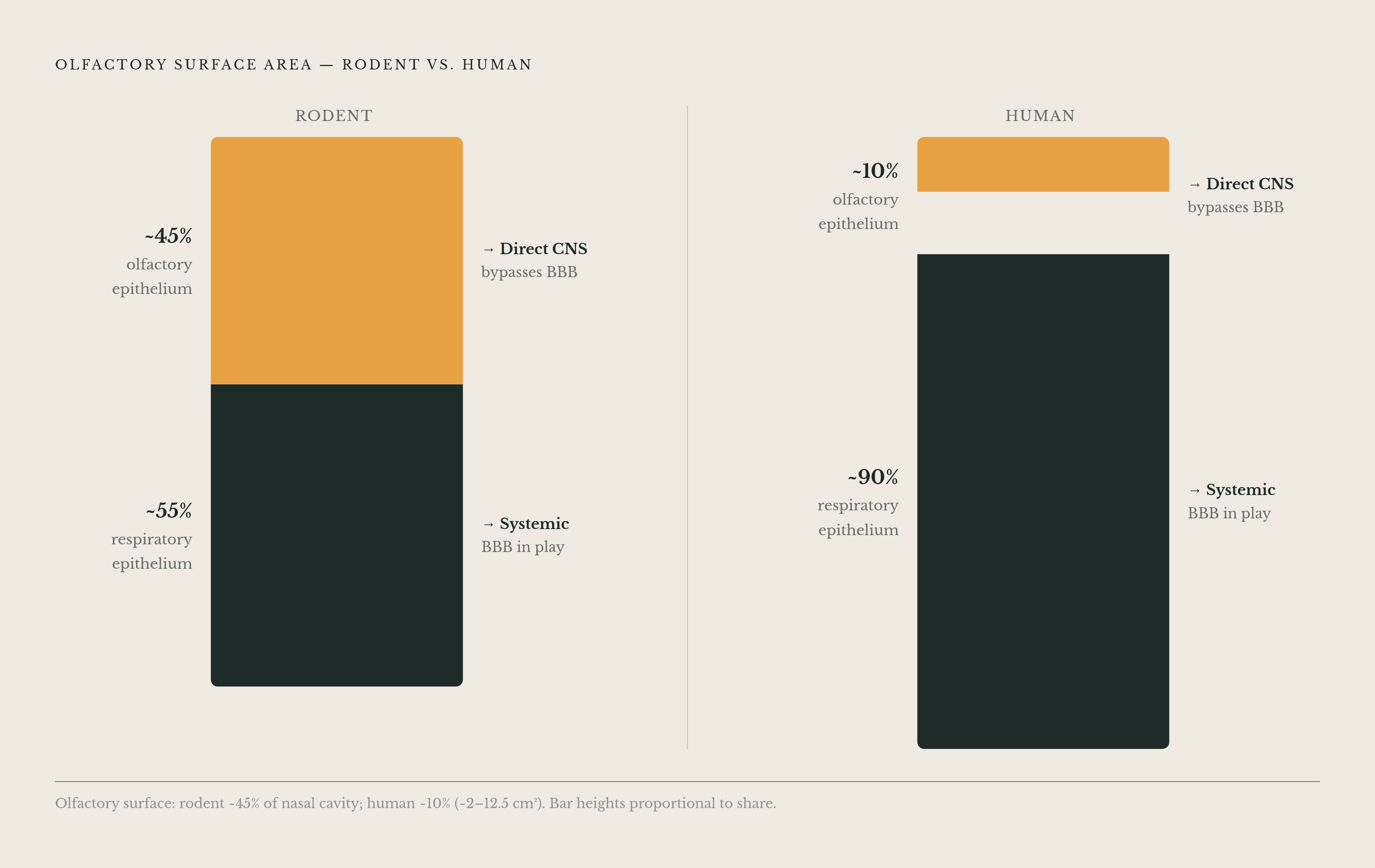
The olfactory epithelium's extraordinary transport capability comes packaged in an anatomically constrained zone. In humans it occupies roughly 10% of total nasal surface area, approximately 2–12.5 cm², tucked into the uppermost recess of the nasal cavity where standard nasal sprays deposit poorly. The structure that enables direct brain transport is also the hardest part of the nose to reach.
The species comparison makes the translational problem concrete. In rodents, where the overwhelming majority of preclinical nose-to-brain data are generated, the olfactory epithelium covers roughly 40–50% of nasal surface area. A formulation tested in a mouse can reach the olfactory zone by default, simply because the zone is large relative to the nasal cavity. The same formulation in a human deposits largely in the respiratory zone, where the direct neural transport pathway is absent. This species difference is the single biggest structural obstacle to translating nose-to-brain peptide delivery from animal models to human therapeutics.
The consequence is not merely quantitative. (2026) name imprecise targeting of the olfactory epithelium as one of the two major unresolved limitations of the field, alongside the lack of standardized in vitro and in vivo models. The anatomical targeting problem is not a solved engineering detail. It is an active research frontier.
One anatomical detail works in the olfactory zone's favor. Less drug is absorbed into the bloodstream and diverted away from the neural pathways. The zone's relative sparseness means that once a drug reaches it, a smaller fraction is lost to systemic clearance.
How formulation design maps onto OE anatomy to exploit each transport pathway
Each structural feature of the olfactory epithelium creates a specific formulation requirement. The mucus layer Bowman's glands produce must be penetrated. The short absorption window imposed by mucociliary clearance, which eliminates most deposited material within 15–20 minutes for unprotected peptides, must be overcome. Particles must be sized to travel perineural spaces or be taken up by olfactory neuron axons. These are not independent design goals. They are constraints imposed by the same anatomy described in the preceding sections.
Nanoparticle size is the clearest example of anatomy directly dictating formulation. For intranasal nose-to-brain delivery, most peptide- or protein-loaded nanocarriers studied fall within the range of 50 to 150 nm. Particles too large for the perineural spaces cannot take the extracellular route. Particles engineered without attention to this constraint will fail regardless of their chemical sophistication.
The trigeminal-versus-olfactory pathway distinction has direct design consequences. One targeting the brainstem via the trigeminal route may prefer a particle that remains intact through transit. The same carrier geometry can be optimal for one pathway and suboptimal for the other.
Cell-penetrating peptides show how formulation can compensate for the surface area constraint. Chemical modification of the delivery vehicle extended the reach of a small target zone. Formulation strategy can overcome both the BBB and the olfactory epithelium's size limitation for peptides that would otherwise be excluded from the brain entirely.
GLP-1 receptors in the brain make precise OE-targeted delivery a neurological priority, not just a metabolic convenience
GLP-1 receptors are expressed at high levels in brain regions including the neocortex and hippocampus. These are the same regions that intranasal administration of high-molecular-weight GLP-1 agonists like dulaglutide has been shown to reach in preclinical studies. The convergence between the delivery route's anatomical reach and the biological targets that matter gives nose-to-brain delivery a pharmacological rationale beyond bypassing the BBB.
The receptor distribution is specific, not diffuse. A 2025 study demonstrated that low-dose semaglutide significantly reduced some measures of alcohol consumption, including drinks per drinking day and heavy drinking days, and weekly alcohol craving in adults with alcohol use disorder, though it did not significantly reduce average drinks per calendar day or overall drinking days. GLP-1 receptor agonists also influence dopamine transporter function within mesolimbic pathways, which connects the receptor biology to reward circuitry directly.
The neurodegeneration evidence points toward prevention rather than treatment. A large 2025 observational study found significantly lower risk of all-cause dementia, Alzheimer's disease, and Parkinson's disease in GLP-1RA users with Type 2 diabetes. The operative window appears to be before clinical diagnosis, when the receptor-mediated pathways can still be modulated.
For Parkinson's disease, the preclinical and clinical picture is more direct. GLP-1 receptor agonists reduce motor symptoms and support dopaminergic function in preclinical models. Clinical trials have shown improved non-motor symptoms and quality of life with liraglutide.
The delivery route matters because the injected route cannot reach the same brain regions efficiently. Peptides with high molecular weight and low lipophilicity, the profile of most GLP-1 receptor agonists, are generally limited in their ability to diffuse across the BBB even after systemic administration. The neurological receptor distribution that makes GLP-1s promising for addiction and dementia is pharmacologically inaccessible by injection in the way it is accessible via the olfactory route. Drath, Richter, and Feja (2025) confirmed that neuropeptides including GLP-1 and GLP-2 derivatives were rapidly transported through trigeminal axons after intranasal application in mice. The pathways described in this piece can carry the specific therapeutic class most relevant to these neurological indications.
Where the science is genuinely unresolved: targeting precision and the preclinical-to-clinical gap
The anatomical argument for the olfactory epithelium as a direct CNS delivery route is well established. What remains unresolved is whether current delivery systems can reliably and precisely target the olfactory epithelium in humans rather than depositing drug in the respiratory zone, and whether brain concentrations seen in rodent models translate to therapeutic levels in human tissue. D'Souza et al. (2026) identify imprecise olfactory epithelium targeting and the lack of standardized in vitro and in vivo models as the two major limitations blocking clinical translation. The field does not yet have agreed-upon tools to measure olfactory-specific delivery or to compare results across studies. That modeling gap is itself a form of imprecision: two studies reporting intranasal brain delivery may be measuring different things if one corrects for systemic contribution and the other does not.
The rodent-to-human surface area gap cannot be closed by formulation chemistry alone. A well-designed formulation that achieves excellent olfactory targeting in mice may deposit largely in the respiratory zone in humans. Device innovation and human anatomical modeling are required to move from animal demonstration to human reliability.
The strongest objection to the field is that despite remarkable preclinical results enhancing brain drug concentrations, clinical translation has lagged. A 2026 expert review published in the Journal of Pharmaceutical Sciences states that this gap remains the central unresolved challenge. The answer to that objection is not that the preclinical results are unreal. It is that the gap is an anatomical targeting and modeling problem, not a disproof of the transport mechanisms the olfactory epithelium's structure enables.
The first-in-human PET imaging study of intranasal oxytocin detected brain uptake between 25 and 45 minutes, but levels were low and variable across individuals. To achieve consistent therapeutic concentrations, you need further optimization of both formulation and delivery device. That optimization is the active engineering frontier, not a problem that has defeated the field.
Sources
- Nose-to-brain drug delivery: from bench to bedside - PMC
- Intranasal drug delivery: Unlocking the nose-to-brain route for central nervous system therapies - ScienceDirect
- Full article: Differential targeting of olfactory epithelium and respiratory epithelium in nose-to-brain drug delivery
- The Nasal–Brain Drug Delivery Route: Mechanisms and Applications to Central Nervous System Diseases