Olfactory Route Peptide Kinetics Versus IV in Deep Brain Structures What the Concentration-Time Curves Actually Show
Intranasal peptides reach the brain differently than IV doses.
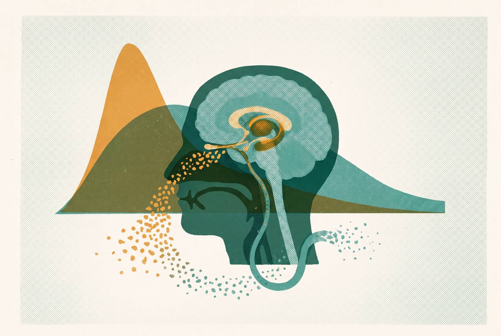
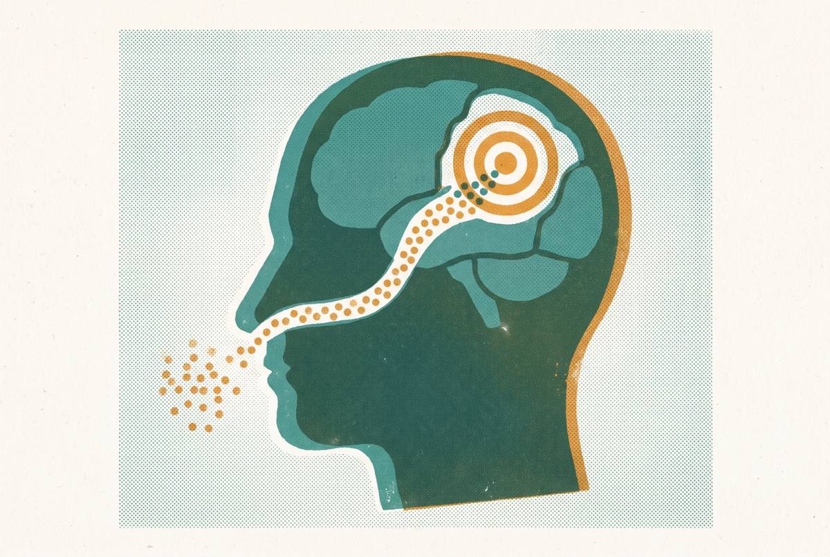
Nanoparticle nose-to-brain delivery for neurological indications uses olfactory and trigeminal pathways to approach the CNS without requiring blood–brain barrier passage. Compared with IV delivery, intranasal peptides can change peak times, regional exposure and brain-to-plasma ratios as well as bioavailability. Those profiles must deliver enough peptide to the intended deep brain structure beyond the olfactory bulb.
Why IV and olfactory-route peptides produce fundamentally different concentration-time profiles in the brain
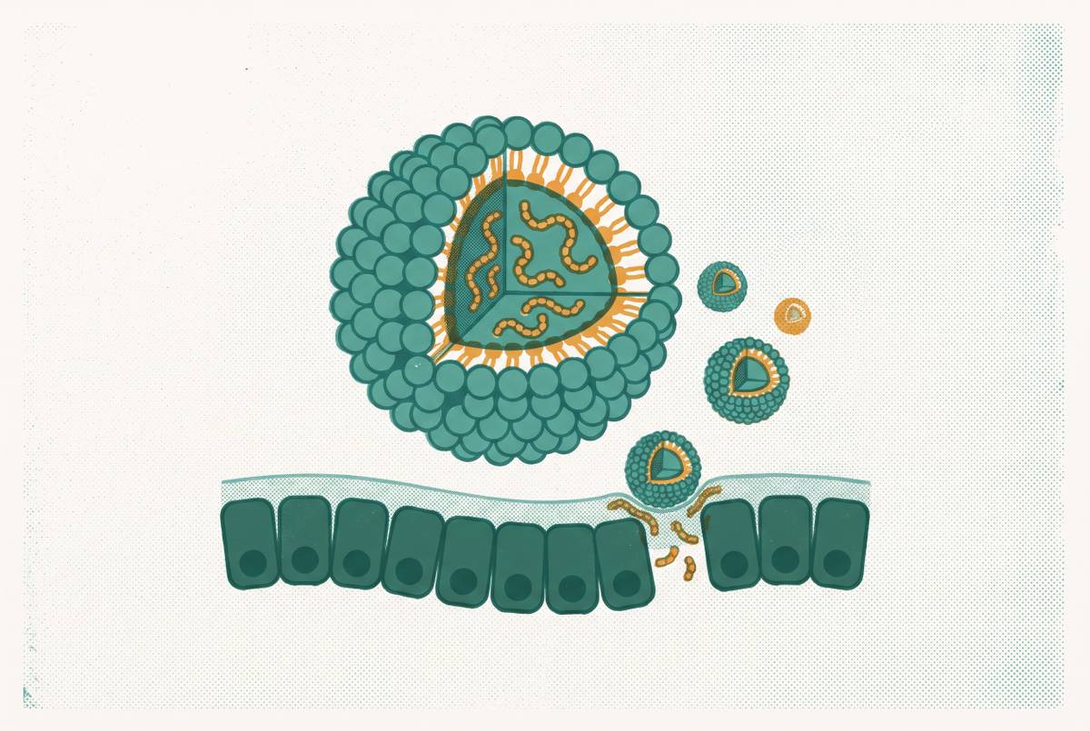
Nanoparticle-encapsulated fibroblast growth factor provides a striking starting point, with an intranasal Direct Transport Index reaching up to 1050% in the olfactory bulb. That is a transport metric, not the percentage of a dose recovered in brain tissue. Does the same advantage reach deep structures?
The assumption that whichever route delivers more drug faster wins cannot answer that question. IV produces a sharp systemic peak followed by decline, while intranasal delivery can produce a lower, later plasma peak and a more favorable brain-to-plasma ratio. Absolute brain concentration can nevertheless be lower after nasal administration. Understanding their relationship requires starting with where each dose enters.
The anatomy that makes the two routes non-equivalent from the first moment of dosing
IV administration places a peptide in the vascular system. To reach brain tissue, it must then cross the blood–brain barrier, a highly selective physical barrier that severely restricts most peptides. The nasal route begins on a mucosal surface connected to two cranial nerve pathways, the olfactory nerve and the trigeminal nerve. Those connections offer access to the CNS without requiring passage across the blood–brain barrier. The entry systems are different from the outset.
The olfactory nerve, cranial nerve I, extends from the olfactory epithelium through the cribriform plate to the olfactory bulb. From there, a peptide can move on toward the hippocampus and cortex. The trigeminal nerve, cranial nerve V, innervates the respiratory epithelium across the broader nasal mucosa and connects to the brainstem through the trigeminal ganglion. The destinations are therefore not interchangeable. An olfactory signal and a brainstem signal need not represent the same delivery path.
Within these routes, molecules may move inside axons, along extracellular perineural spaces or through supporting cells. The perineural spaces connect directly to cerebrospinal fluid in the subarachnoid space. Access to those spaces also depends on nasal geometry. In humans, the olfactory epithelium occupies less than ten percent of nasal surface area, tucked into an upper recess where standard sprays deposit poorly, whereas rodents have proportionally much greater coverage. This species difference limits translation before transport speed is considered.
The olfactory region has lower vascular density than the respiratory region, which correlates with higher brain delivery efficiency because less drug is diverted into blood. Systemic absorption is therefore a competing destination. Nasal deposition cannot be treated as a single anatomical event when different surfaces connect the dose to different routes.
How transport mechanism shapes the rising phase of the intranasal concentration-time curve
Once a peptide reaches a suitable surface, how it mainly travels shapes the rising curve. Extracellular perineural transport operates within minutes, while intracellular axonal transport can take hours to days. Transcellular transport through supporting cells is another way across the local tissue. A rapid signal from released drug is not equivalent to rapid movement of an intact nanoparticle.
Intranasal phenytoin can reach maximum CSF concentration 5 minutes after application, demonstrating how quickly access to that compartment can occur. That observation does not establish the same timing for a peptide or for deep brain tissue. For free, unmodified peptides, concentration gradients drive transcellular or paracellular diffusion along olfactory and trigeminal pathways. Enzymatic degradation in the nasal mucosa is the principal loss mechanism that blunts this rise.
Nanoparticle encapsulation changes which material moves along which pathway. The trigeminal route serves as the dominant pathway for intact nanoparticles, while the olfactory route is more likely to deliver substances already released from them. Where trigeminal axonal transit governs larger encapsulated peptides, the expected rise is slower and more sustained. Formulation therefore helps determine the slope itself.
Cell-penetrating peptide conjugation and mucoadhesive polymers provide further ways to extend the absorption window and shift transport. Their purpose is not necessarily to create the earliest possible peak. Mucociliary clearance also limits the time available for absorption, so residence time affects absorption kinetics. Which process dominates depends on the molecule and formulation. “Intranasal” consequently describes a family of possible curves.
What the head-to-head concentration-time data from deep brain structures actually show
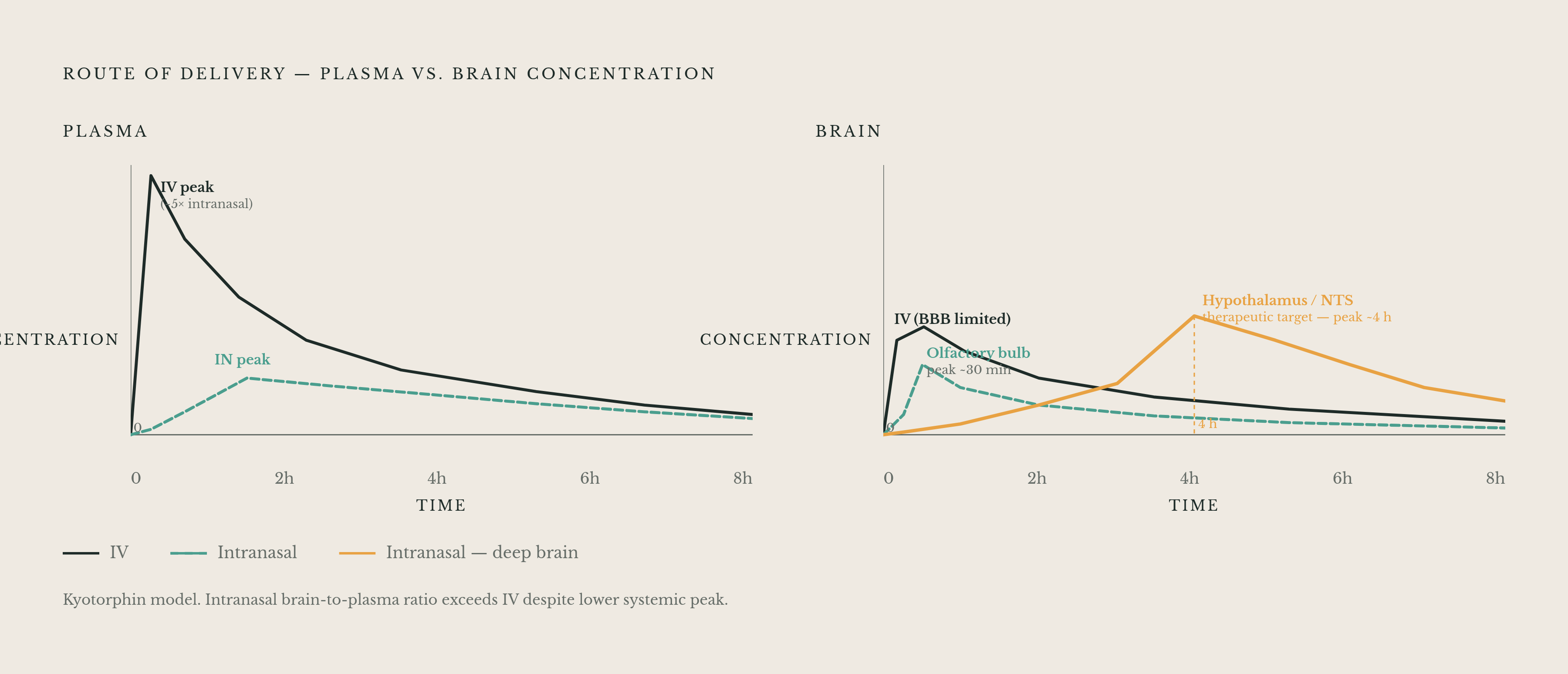
The routes differed in how much kyotorphin reached the plasma and when. After intranasal administration, blood concentration rose slowly and plasma peaked at 30 minutes, while IV administration led to a rapid peak and then a steady decline. The peak with IV was roughly five times higher. Yet the brain-to-plasma ratio was higher with intranasal delivery even though absolute brain concentration was lower.
The fibroblast growth factor comparison answers a different question. Intranasal bFGF nanoparticles achieved higher brain area under the concentration-time curve, or AUC, than IV administration. Direct Transport Efficiency and the olfactory-bulb transport index supported delivery straight to the brain over redistribution through the systemic circulation. These findings establish a stronger exposure result for this formulation, but they do not erase the distinction between whole-brain measurements and regional concentrations.
The regional data expose that distinction directly. Peak concentration in the rest of the rat brain was not reached until several hours after application. IV produces a different entry-point gradient because systemic distribution brings drug to vascularized tissues at roughly the same time, with brain entry depending on blood–brain barrier permeability. The two routes impose different spatial sequences on the concentration-time profile, with IV exposure also varying across brain structures.
Winterdahl’s 2025 PET study provides the first-in-human imaging anchor in this evidence. It found brain uptake within tens of minutes, but uptake was low and differed from person to person. Human imaging supports the existence of the route without establishing dependable, therapeutically sufficient exposure in a particular deep structure. Whether the rodent regional profile translates intact remains unresolved.
Why GLP-1 receptors in the hypothalamus and brainstem make the regional gradient the clinically decisive variable
For GLP-1 delivery, the relevant receptor map extends well beyond the olfactory bulb. Hypothalamic GLP-1 receptors occur in the arcuate, paraventricular and dorsomedial nuclei, as well as lateral hypothalamic areas involved in feeding and energy balance. Brainstem sites include the nucleus tractus solitarius, or NTS, the area postrema and the dorsal motor nucleus. A delayed deep-brain peak therefore affects the very regions that the treatment aims to reach.
The circuitry explains why those destinations are important. Gut-secreted GLP-1 modulates vagal pathways whose signals reach the NTS and are relayed to hypothalamic nuclei. Centrally secreted GLP-1 from the NTS projects widely to align food intake with energy demands, while most preproglucagon neurons lie in the lower brainstem and project toward forebrain areas. The brainstem system also reduces motivation and reward-seeking behavior. Elsewhere, GLP-1 receptor activation promotes hippocampal neurogenesis and modulates dopaminergic and serotonergic pathways involved in mood and reward.
Preclinical subcutaneous studies show very limited brain penetration, with brain-to-plasma ratios far below one. MetP Pharma AG, based in Switzerland, reported in January 2026 that its intranasal semaglutide technology produced ratios consistently greater than one in rats. This was a company-reported preclinical result. It does not by itself identify exposure at the hypothalamic nuclei or NTS. If the bulb peaks at 30 minutes while the deep structures of interest peak around 4 hours later, does enough peptide reach those targets, or does most of the apparent gain stay at the olfactory relay station?
The honest reckoning with the olfactory-to-deep-brain gradient at the hypothalamus and NTS
Olfactory-bulb accessibility does not establish hypothalamic or brainstem delivery. For GLP-1 analogues specifically, attenuation between the bulb and deep hypothalamus remains uncharacterized in peer-reviewed human data. The supplied findings also lack peer-reviewed data on human exposure at the NTS. This is the central evidence gap. It concerns the amount of peptide reaching its target, the time required and the persistence of exposure there.
The exact nose-to-brain transport mechanisms also remain incompletely understood, despite evidence that the route functions in animals and humans. The olfactory corridor takes up a larger share of the nose in rodents than in humans, so good delivery in rodents cannot show whether the drug reaches its targets in humans. The PET finding makes that uncertainty concrete. Detectable uptake is compatible with low exposure and substantial individual variation. Neither rapid CSF access nor a whole-brain ratio resolves the missing regional measurement.
Eliasson et al.’s January 2026 bioRxiv study from Lund University and Trulylabs examined a device-based lever through NosaPlugs nasal inserts. It demonstrated cumulative memantine release over an extended period in vitro and pharmacologically relevant plasma and brain levels in mice. Retention time is therefore a plausible engineering lever. Evidence of GLP-1 analogue delivery to the human hypothalamus or NTS remains needed beyond this memantine study.
Nanoparticle encapsulation, cell-penetrating peptide conjugation and mucoadhesive polymers can extend the absorption window and alter transport, but none closes the human regional evidence gap presented here. The obstacle leaves the route viable in principle but requires a credible platform to confront regional attenuation and delayed arrival through deep-brain pharmacokinetics beyond a bulb measurement.
How should nanoparticle nose-to-brain delivery platforms for neurological indications be designed?
Where deep structures peak hours after the bulb, formulations must extend mucosal residence and sustain release rather than simply maximize the initial deposition peak. A high early nasal or olfactory signal answers the wrong timing question if exposure disappears before the intended destination is adequately supplied. Regional selectivity changes the specification. An entry-point advantage may suit olfactory-bulb or hippocampal targeting but demands further transport characterization for the arcuate nucleus or NTS.
The optimization target should include both the CNS-to-plasma ratio and absolute brain concentration. A favorable ratio shows brain exposure with less systemic exposure alongside it, which gives a reason to try this route to reduce peripheral side effects. It does not establish adequate target concentration by itself. For injectable GLP-1 therapies, systemic exposure is associated with gastrointestinal adverse effects and nausea that can drive discontinuation.
The neurological opportunities remain mechanistic hypotheses awaiting demonstration through intranasal human outcomes. GLP-1 receptor agonists may increase brain glucose metabolism and help glucose cross the blood–brain barrier, both of which are relevant to Alzheimer’s pathology. The brainstem system’s role in motivation and reward makes the NTS a logical addiction target, approached anatomically through the trigeminal connection to the brainstem. Each hypothesis depends on meaningful local exposure. A route to a region is not yet a therapeutic concentration within it.
The Parkinson’s clinical record reinforces the need to separate mechanism from outcome. LixiPark showed potential disease-modifying effects, while the EXENATIDE-PD3 phase 3 trial found no evidence supporting exenatide as disease-modifying. Differences in CNS exposure may contribute to such outcomes, but these results do not establish that explanation. Nanoparticle platforms with peer-reviewed pharmacokinetic data for other CNS molecules provide a foundation for testing delivery, with GLP-1 efficacy still to be established. The concentration-time curve is the engineering map, showing where the route is fast, where it is slow and which formulation decisions can change the journey.
Sources
- Nose-to-brain drug delivery: from bench to bedside
- First In Vivo Demonstration of Nose-to-Brain Drug Delivery ...
- Advanced intranasal peptide delivery systems for improved management of Alzheimer's disease - ScienceDirect
- Editorial: Intranasal Delivery of Central Nervous System Active Drugs: Opportunities and Challenges
- Investigation of the Transport Pathways Associated with Enhanced Brain Delivery of Peptide Drugs by Intranasal Coadministration with Penetratin
- Glucagon-Like Peptide-1 and Hypothalamic Regulation of Satiation: Cognitive and Neural Insights from Human and Animal Studies
- Brain-Derived GLP-1—Understanding the Physiological Function and Anti-obesity Potential of Preproglucagon Neurons